Cornell researchers create faster imaging method to track cell metabolism
Cornell University scientists have developed a new technique for real-time observation of cell metabolism that was published on September 4.
New tool speeds up screening
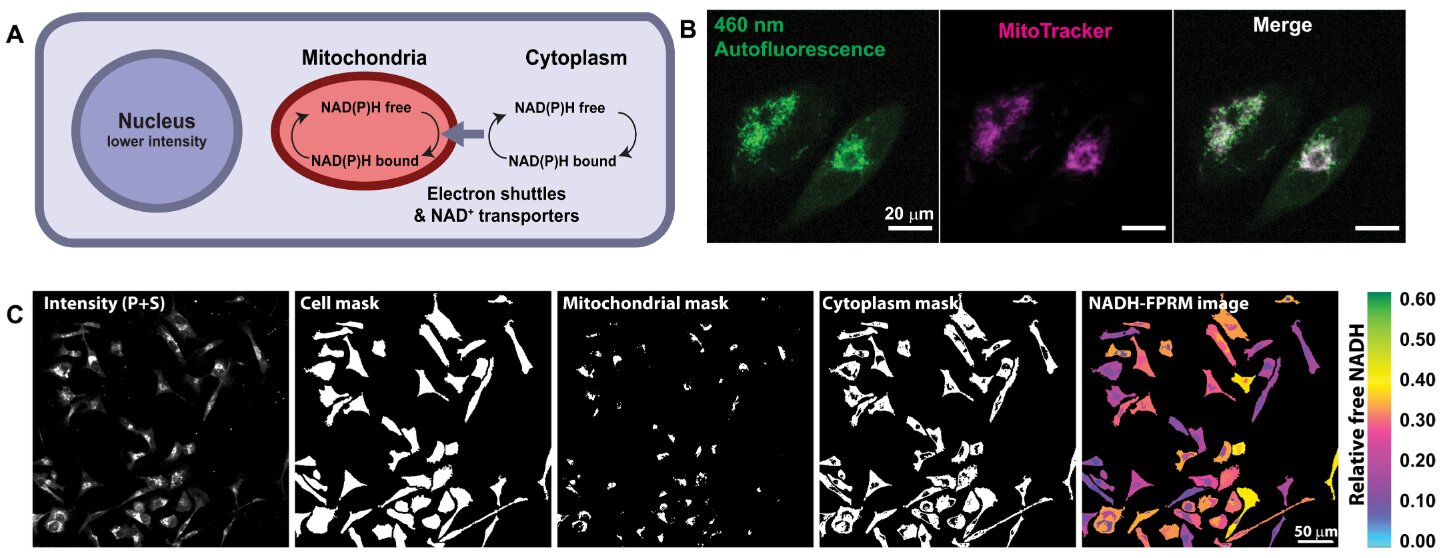
The team designed a faster version of two-photon fluorescence imaging to observe metabolic changes in cells. This approach allows researchers to screen new therapeutic treatments much more quickly than previous methods could.
Dynamic processes require better tools
Lu Ling started the project because standard tools could not track rapid changes happening within minutes or hours. She noted that cancer metabolism is a very dynamic process requiring improved observation capabilities.
Collaboration brings new expertise
Researchers contacted Warren Zipfel to utilize his specialization in optical microscopy for biomedical research. The group also focused on fluorescence lifetime imaging microscopy which monitors metabolism based on NADH fluorescence.
Reported by one outlet
Only one outlet has published this. Nothing here has been checked against a second report, so read it as that outlet's account and follow the link below for the original.
Reported by
1 independent outlet. Headline as published. Links open the original report.