Researchers use atomic force microscopy to study antibody access on cancer vesicles
Scientists at Kanazawa University have developed a method using high-speed atomic force microscopy to observe how antibodies interact with cancer-related proteins on tiny vesicles.
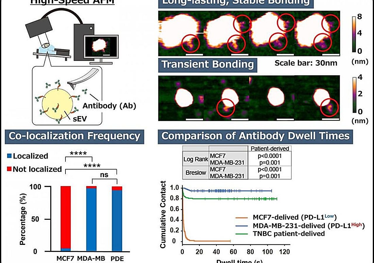
New imaging technique reveals protein accessibility
Researchers found that measuring the total amount of a protein does not always show how accessible it is to antibodies. Some proteins may be present but hidden by nearby molecules or positioned in ways that block antibody reach. This physical availability is known as accessibility and differs from simple quantity measurements.
Study focuses on PD-L1 interaction
The investigation centers on programmed death-ligand 1, a protein that can act as a brake on immune cell attacks against tumors. Medicines designed to block this interaction have changed cancer treatment outcomes, yet patient responses vary significantly. Current tests often measure how much of the protein exists without accounting for its physical position.
Findings published in Small Methods
The team reported these results in the journal Small Methods after using high-speed atomic force microscopy for real-time observation. Further research is required before this approach can be applied in clinical settings to help patients. The new method allows investigation of differences between individual cancer-derived vesicles released by cells.
Reported by one outlet
Only one outlet has published this. Nothing here has been checked against a second report, so read it as that outlet's account and follow the link below for the original.
Reported by
1 independent outlet. Headline as published. Links open the original report.