Scientists use cryo-electron tomography to visualize bacterial protein coordination
Researchers from EMBL Heidelberg published findings in Cell about how bacteria coordinate protein production and transport using new imaging tools.
New imaging technique reveals cell details
Scientists have used cryo-electron tomography to visualize connections between different cellular processes inside flash-frozen cells. This method allows researchers to see structures in three dimensions with great detail rather than looking at molecules in isolation.
Study focuses on specific bacteria type
The team imaged Mycoplasma pneumoniae bacteria to examine interactions between various cellular systems. Joe Dobbs, a lead author and former Ph.D. fellow in the group, noted that both papers serve as proofs of concept for this analysis.
Findings published in scientific journal
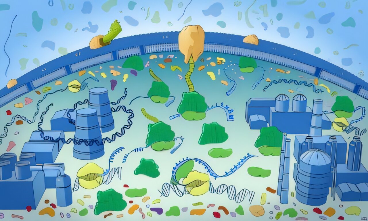
The latest research results appear in two separate papers within the journal Cell. These studies help explain how bacterial cells manage shipping systems and factories within their small, bustling city-like structures.
Reported by one outlet
Only one outlet has published this. Nothing here has been checked against a second report, so read it as that outlet's account and follow the link below for the original.
Reported by
1 independent outlet. Headline as published. Links open the original report.