Researchers at Uppsala University reveal how cancer drugs differ in 3D cell clusters
Scientists at Uppsala University used transparent minispheres to show that two similar cancer drugs affect bowel cancer cells differently when grown in three dimensions.
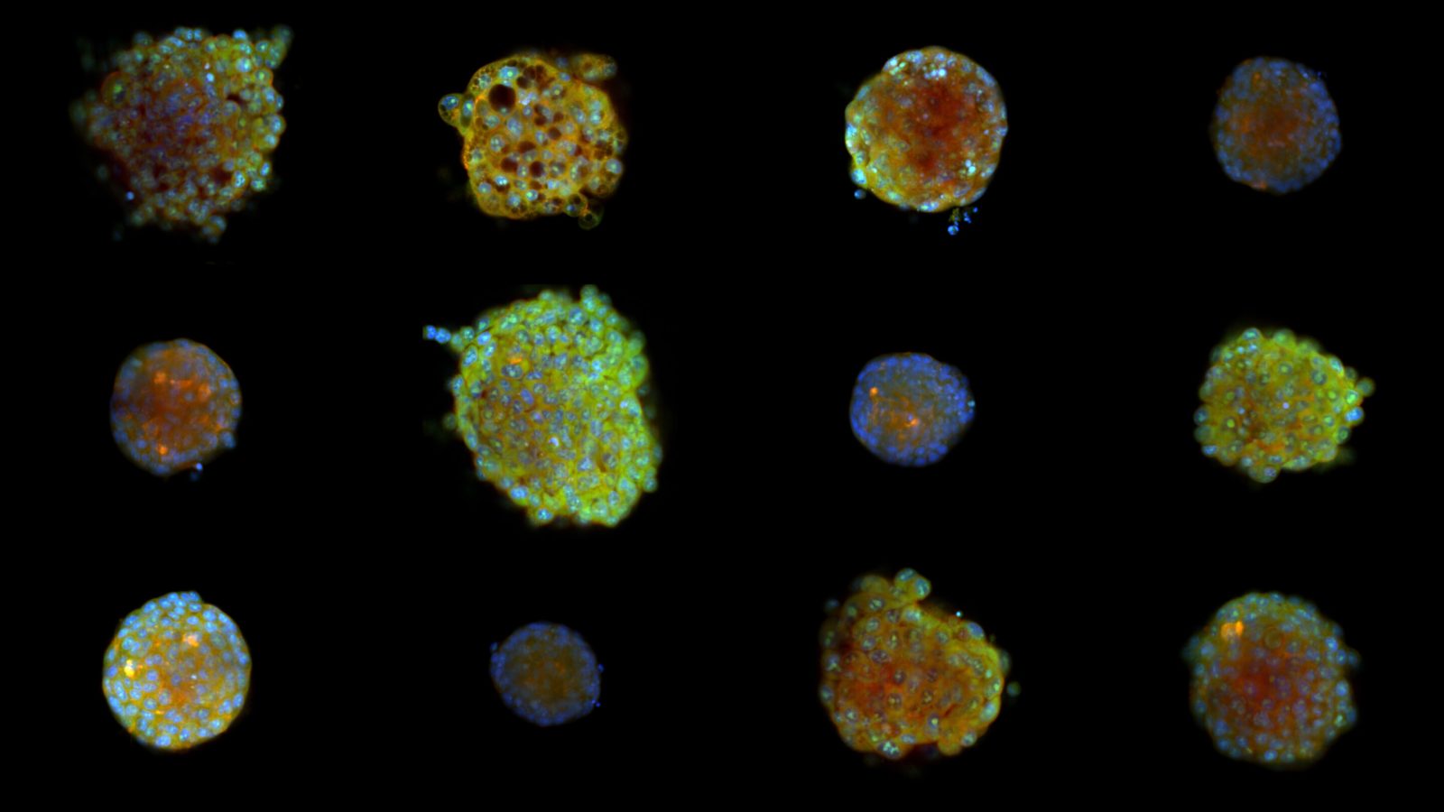
New imaging method for 3D clusters
Researchers adapted the Cell Painting imaging method to examine individual cells inside small 3D clusters of cancer tissue. They made these minispheres transparent using a sugar and glycerol solution so microscopes could see deep inside them. This approach allows scientists to measure thousands of details about each cell's shape and structure without collapsing the sample.
Drugs behave differently in 3D
Two drugs that appeared almost identical in flat cultures showed distinct behaviors when tested inside these 3D spheres. The team linked this difference to how each drug activates the cell's p53 stress response mechanism. Most screening methods currently use flat layers or flatten entire samples, which hides this important variation.
Study published in Nature Communications
The work has been published in the journal Nature Communications by researchers from Uppsala University. The study highlights that cells behave differently when they grow in three dimensions compared to flat layers. Drug developers can now use this technique to see how treatments change cells in a more realistic environment.
Reported by one outlet
Only one outlet has published this. Nothing here has been checked against a second report, so read it as that outlet's account and follow the link below for the original.
Reported by
1 independent outlet. Headline as published. Links open the original report.